- shafiedarou
- August 29, 2023
- دسته بندی نشده
- 0 Comments
For medical professionals such as radiologists and physicians to comprehend what’s happening inside our bodies, they may ask patients to undergo contrast media treatment. Contrast media is used in radiology procedures for delineating tissues with poor inherent contrast to help diagnose issues by highlighting internal areas and structures of the body. Also referred to as “contrast materials” or “contrast agents”, contrast media are liquid substances which temporarily color areas of the body to improve the diagnostic effectiveness during imaging procedures.
They were initially utilized to better visualize vascular structures and the gastrointestinal tract. Over time they become one of the key components of medical imaging and their use extended to the evaluation of vascularization and perfusion of tissue in solid organs, specifically using cross-sectional imaging techniques and enhanced visualization and characterization of focal lesions within these organs. The use of contrast agents is indispensable in radiology, and it will remain as such in the foreseeable future.
Contrast media is a valuable imagining solution applied across the following medical imaging exams:

Specific contrast media will use certain chemical combinations depending on the procedure. For example, iodine and barium-based compounds support X-ray and CT exams while gadolinium is for MRI and microbubbles are for ultrasound procedures.
Contrast Media are classified based on different factors such as Modality, Type, Route of Administration, and Indication.
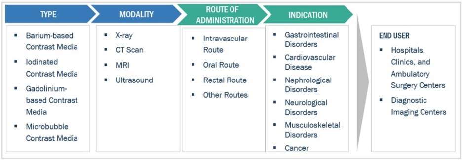
In X-ray and CT exams, there are many ways for categorizing contrast media:
- Atomic weight: Under this classification we have the positive and negative contrast media. Positive contrast media has its name because it possesses a higher atomic weight than surrounding tissue. This means that it will absorb more photons than the tissue that surrounds it and would produce a lower radiographic density than them. E.g. Barium sulphate, Organic Iodine. Negative contrast media: Has a lower atomic weight than surrounding tissue that it would absorb less photons and would produce greater radiographic density than the tissues surrounding it. e.g. air, oxygen, carbon dioxide.
- Solubility: The positive contrast media barium sulfate and organic iodine are further classified based on their solubility in water. Barium sulfate is a water insoluble contrast agent so it is not readily absorbed and excreted by the body. This means that it will stay within the body cavity for a long period of time. Barium sulfate usually produced in a powder or semisolid suspension like a syrup. These contrast media used majority in investigations of the gastrointestinal tract. Depending on the examination the barium sulfate is either taken orally or through the rectum as an enema.
Water soluble contrast media: The organic iodines fall under this group. This type of contrast media usually exists in a liquid form that is soluble in water. When it is introduced into the body it is rapidly absorbed into the body’s water and excreted by the kidneys. Thus, it does not stay within the region of interest for long. This is why speed is important in investigations where this type of contrast media is used. This type of contrast media is used in investigations of the urinary, biliary, cardiovascular, and central nervous systems.
- Osmolarity: Organic iodines are further classified into High Osmolar Contrast Media [HOCM] and Low Osmolar Contrast Media [LOCM]. Osmolality is the concentration of dissolved particles in a solution. A solution with a high osmolality will induce a greater osmotic pressure. This osmotic pressure causes more fluid to flow into the solution. When contrast media with an osmolality far greater than that of body fluid is introduced to the body, the osmotic pressure causes water to move from the low osmolar body fluid to high osmolar contrast media. This leads to dehydration and increased likelihood of intolerance to the contrast media. If contrast media that has an osmolality near the osmolality of the body fluid is used there is a low chance of contrast media reactions occurring. Compared to when contrast media with osmolality far greater than the body fluid is used which has a higher chance of leading to contrast media reactions.
High Osmolar Contrast Media were the first type of organic iodine contrast media to be produced. They are currently the less expensive type on the market. When high osmolar contrast media is introduced into a solution it breaks down into an anion and a cation. The anion is usually iodine while the cation is either sodium or meglumine. This breaking down or dissociation into ions gives the name ionic contrast media. This dissociation into two causes more particles to be present in this type of contrast. As a matter of fact, high osmolarity contrast media have an osmolarity that is five to eight times greater than the body fluid plasma. This type of contrast media has a higher chance of causing a contrast media reaction this is why it has been replaced by low osmolar contrast media in many radiographic examinations. Low Osmolar Contrast Media are advancement over HOCM and are currently more expensive than the high osmolar contrast media.
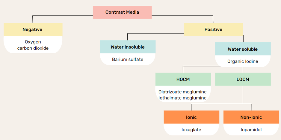
A summary of the types of contrast Media classification in X-ray and CT Procedures
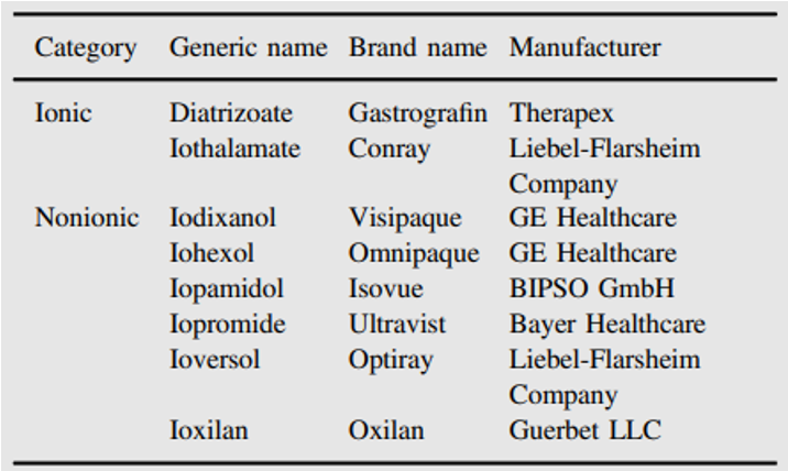
Some commercially available small molecule iodinated contrast media are listed in below.
Magnetic Resonance Imaging (MRI) Contrast agents:
Magnetic resonance imaging (MRI) contrast agents are categorized according to the following specific features:
- chemical composition including the presence or absence of metal atoms
- route of administration, magnetic properties
- effect on the magnetic resonance image
- biodistribution
- imaging applications
The majority of these agents are either paramagnetic ion complexes or superparamagnetic magnetite particles and contain lanthanide elements such as gadolinium (Gd3+) or transition metal manganese (Mn2+).
Gadolinium based Contrast Agents (GBCAs):
Regarding the chelate type and charge, GBCAs can be divided into linear/macrocyclic and ionic/non-ionic groups. A wide range of GBCAs are commercially available and their stability is dependent on the conditional thermodynamic stability constant (Kcond), thermodynamic stability constant (Ktherm), and kinetic stability.
Multiple studies show that the brain holds onto more gadolinium particles from linear agents than macrocyclic agents. Early studies show that most macrocyclic molecules filter through the kidneys and leave the body within 24 hours after an MRI. The rest exit the body within 72 hours.
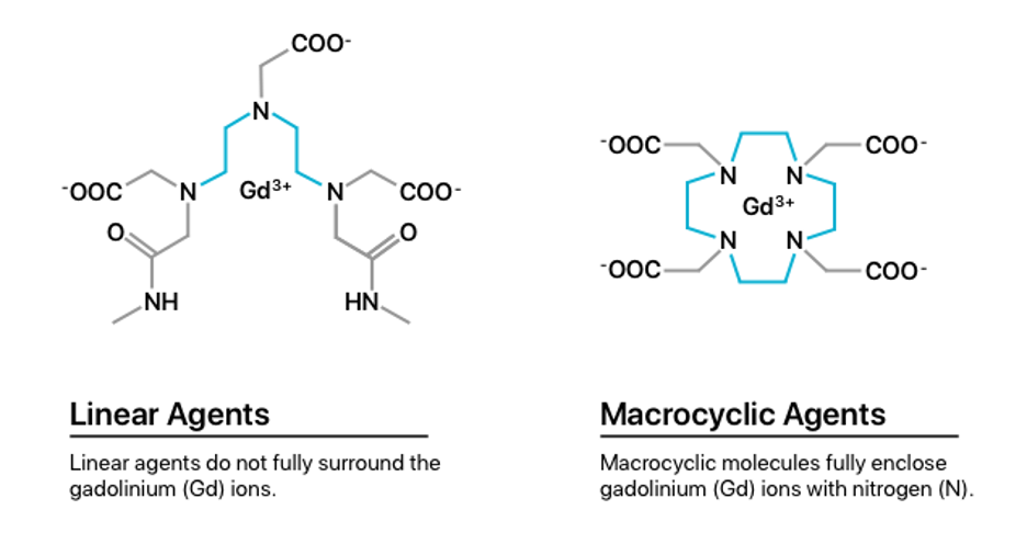
Ionic GBCAs are more stable than nonionic ones and the stability of macrocyclic compounds is higher than linear compounds. Hence, ionic macrocyclic agents are the most stable Gd chelates. Macrocyclic molecules bind strongly to Gd in an organized rigid ring; however, linear nonionic GBCAs have open chains and weaker binding to Gd. Compared to linear agents, macrocyclic agents are more stable in vivo. Low-stability GBCAs (linear, nonionic compounds) likely undergo transmetallation, release free Gd that deposits in tissues, attract fibrocytes, and therefore initiate the process of fibrosis.

MultiHance (gadobenate) is a gadolinium based MRI contrast agent that has twice the relaxivity of conventional extracellular fluid (ECF) contrast agents, providing a marked increase in SNR to better visualize smaller lesions and improve the delineation of larger lesions.

Magnevist (gadopentetate dimeglumine) Injection is a contrast agent that is used in combination with MRI to allow blood vessels, organs, and other non-bony tissues to be seen more clearly on the MRI used to help diagnose certain disorders of the heart, brain, blood vessels, and spinal tissues.

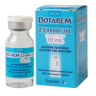
Dotarem (Gadoterate meglumine also known as gadoteric acid) is a contrast agent that has magnetic properties. It is used in combination with MRI to allow blood vessels, organs, and other non-bony tissues to be seen more clearly on the MRI. Dotarem is used to help diagnose certain disorders of the brain and spine (central nervous system).
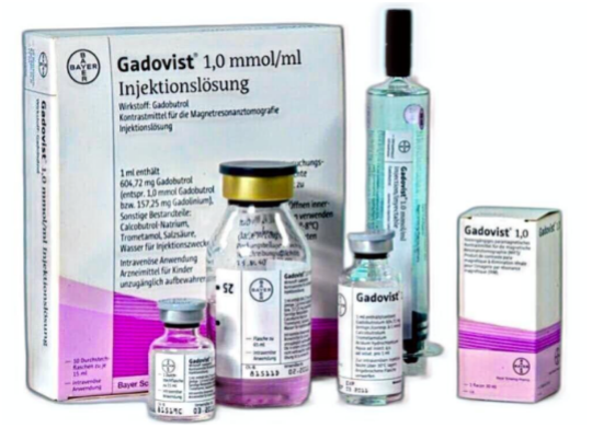
Gadovist (gadobutrol) injection is indicated in adults and children of all ages including term newborns for: Contrast enhancement during cranial and spinal MRI investigations and for contrast-enhanced magnetic resonance angiography (CE-MRA), Contrast enhanced MRI of the breast to assess the presence and extent of malignant breast disease, and MRI of the kidney.
Side effects of GBCA:Rarely, certain types of gadolinium contrast cause a severe disease called nephrogenic systemic fibrosis (NSF) in people with significant kidney dysfunction. This condition, which causes tightening of the skin and damage to internal organs, is most likely to occur in people with MS who also have kidney dysfunction. Although rare, some people have an allergic reaction to gadolinium contrast. The main symptom is itchy skin, but rashes have also been reported. A life-threatening allergic reaction, known as anaphylaxis, is also possible but unlikely.
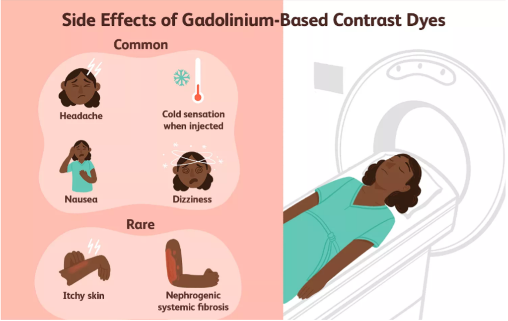
NSF occurs in patients who have kidneys that are not functioning efficiently. When gadolinium is in the bloodstream for longer than is recommended, it begins to deposit in the skin and joints. Symptoms of NSF include a fibrous feel to the skin, limited movement of joints, and muscle tissue that becomes striated.
The first case of NSF was documented in 1997. A link was quickly made between injections of IV contrast in MRI and renal impairment. This disorder can be very serious if it affects the muscles of the lungs, the diaphragm, and the heart.

Key Players:
Some of the prominent players operating in the Contrast Agents in MRI market are:
- Unijules Life Sciences Ltd (India)
- Spago Nanomedical AB (Sweden)
- Lantheus Medical Imaging Inc (U.S.)
- JB. Chemicals and Pharmaceuticals Limited (India)
- Guerbet LLC (France)
- GE Healthcare (U.S.)
- Daiichi Sankyo Company Limited (Japan)
- Bracco Imaging S.p.A (Italy)
- Bayer Healthcare AG (Germany)
- Jodas Expoim (India)
Contrast-Enhanced Ultrasounds (CEUSs)
Ultrasound imaging is a well-established clinical tool for the morphological assessment of soft tissues, employed frequently in obstetrics, cardiology, and radiology. As an ultrasonic wave (which is a longitudinal wave) is transmitted into the body, reflections are generated from tissue interfaces that are characterized by different acoustic properties, i.e., speed of sound and density. These scattered signals are recorded by the same transmitting transducer and used to generate an image.
CEUSs are microbubbles of gas with a protein, lipid or polymer shell. They are blood pool agents and remain confined to the intravascular space after intravenous injection. They are unable to pass through the vascular endothelium. Small microbubbles (1-10 mcm) can pass through the lung capillaries and remain in circulation for a short time and eventually get dissolved. The gas gets exhaled while the shell gets metabolized in the liver.

An important consideration in understanding bubble behavior in CEUS is that these phenomena occur in different proportions at different incident acoustic pressures. Acoustic response of an ultrasound contrast agent is specific for the microbubbles used and also depends on the acoustic power of the irradiated ultrasound wave.
At low acoustic powers, the microbubbles act as simple reflectors, so that only a backscattered linear signal can be received. At intermediate acoustic powers, the microbubbles are induced to oscillate and thereby to emit an intensive non-linear resonance signal, which contains, in addition to the fundamental vibration frequency, also harmonic upper frequencies. At even higher acoustic powers, the microbubble vibration is so violent that the microbubbles are destroyed by tearing of the membranes. This process is accompanied by emission of a detectable ultrasound pulse.

Definity:
Each contrast agent has specific storage and reconstitution instructions that can be found in their respective package inserts. Commercial agents are packaged as single-use vials. Definity one of the most commonly used contrast agent is supplied as a clear liquid in a vial containing approximately 1.5 ml of contrast agent. It’s activated by vigorously shaking it on a special shaker called vial mix for approximately 45 seconds producing this milky substance which is a suspension of microbubbles this suspension is ready to inject surprise stable for approximately five to six hours and can be used throughout normal work day.

Optison:
Optson is supplied as ready suspension of microbubbles it’s agitated by gently rotating vial for approximately three minutes and then ready for injection. Same as other agents it has couple hours shelf life once agitated and can be used in a different patient if needed. The 3-mL vial is stored in a refrigerator at 2-8 °C, and brought to room temperature before use. The vial is inverted and gently rotated until it is a milky white suspension, then vented with either a vent spike or 18-G needle before being withdrawn into the injection syringe.

Sonovue/lumason:
Sonovue is supplied differently. It comes as a dry powder of microbubbles. It comes in a kit with a pre-filled 5 ml syringe which is used to reconstitute some of your microbubbles through a specially provided pin. This pin prevents bursting of the microbubbles while transferring microbubbles from the vial back into the syringe. The kit is stored at room temperature, 25 °C with a permitted range of 15-30 °C. All components needed for reconstitution are within the kit. A vial may be used for up to 6 h from the time of reconstitution.

Key players:
The major players in Ultrasound contrast media market are GE Healthcare (US), Bracco Imaging SpA (Italy, and Lantheus Medical Imaging, Inc. (US).
https://link.springer.com/chapter/10.1007/978-3-030-79256-5_1
https://epos.myesr.org/poster/esr/ecr2019/C-3366/Background#poster
https://www.marketsandmarkets.com/Market-Reports/contrast-media-market-911.html
https://www.drugs.com/pro/gastrografin.html
https://www.gehealthcare.com/products/contrast-media/visipaque
https://pubmed.ncbi.nlm.nih.gov/27666161/
https://www.medical-professionals.com/en/mri-safety-gadolinium-side-effects/
https://www.bracco.com/en-ca/product/isovue
https://www.frontiersin.org/journals/physics/articles/10.3389/fphy.2022.791145/full
https://www.slideshare.net/slideshow/ultrasound-contrast-agent-ppt/259230788
https://www.sciencedirect.com/science/article/pii/S0169409X20300818