- shafiedarou
- August 31, 2023
- دسته بندی نشده
- 0 Comments
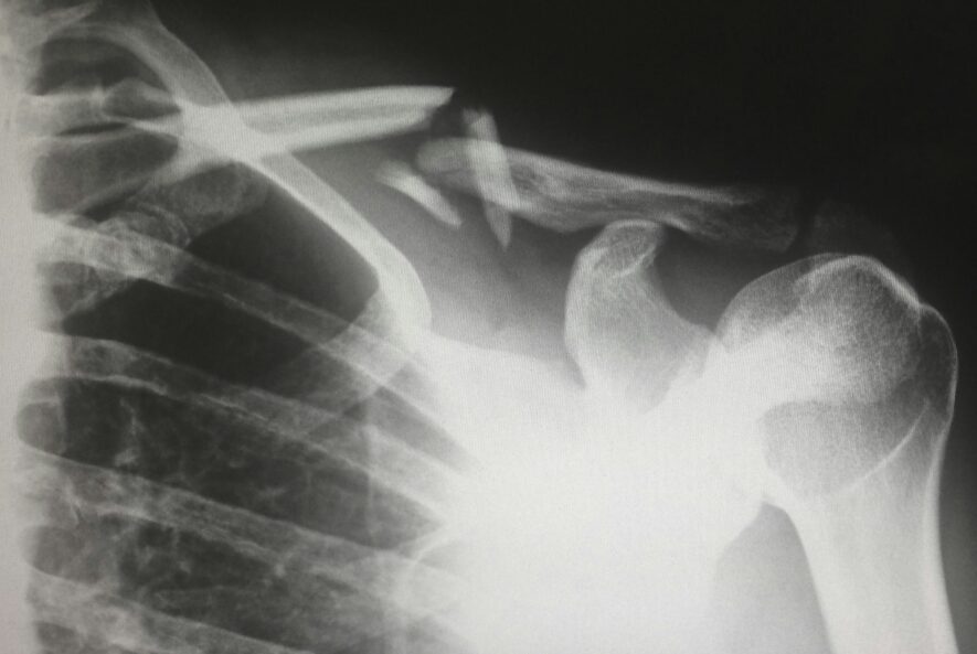
magnetic resonance imaging (MRI) has become widely used in hospitals around the world, since it received FDA approval for clinical use in 1985. It is a clinical imaging modality, which relies on the detection of NMR signals emitted by hydrogen protons in the body placed in a magnetic field. Generally, MRI is used less commonly than plain films and CT scans. They are often reserved for superior viewing of soft tissues. MRI is particularly helpful in patients with suspected neurological or musculoskeletal pathology; however, they can be used in many other specialities too. It takes slightly longer to acquire MR images and they are more expensive. MRI is contraindicated in patients who have ferromagnetic metal implants or foreign bodies.
T1 and T2 Weighted Images:
Human body is made up of water, which means a large number of atoms inside our body is hydrogen atoms, the nucleus of which contains a positively charged proton that spins around an axis. Protons behave like small magnets spinning on its randomly aligns axis.

So, what MRI scanners actually do they create a magnetic field. In MRI strong magnets produced magnetic fields that forces all proton axis in the body to align with it; before this field is presence protons are pointing
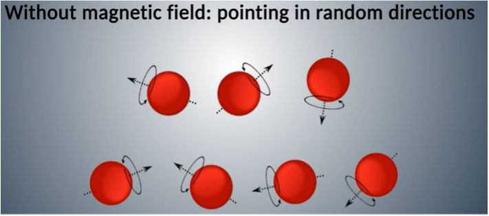
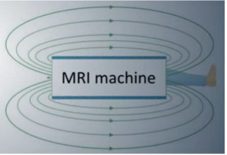
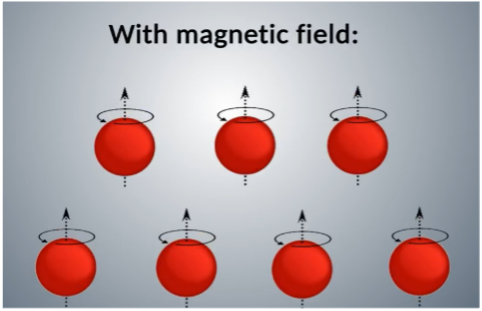
in random directions. When the proton axis is aligned with the fields a radiofrequency current imposed through the patient’s stimulation of the protons will lead to a spin out of equilibrium against the pool of the magnetic fields.
When the RF pulse stops:
- Absorbed RF energy is released by protons
- T1 relaxation (recovery): recovery of longitudinal orientation; T1 time refers to interval where 63% of longitudinal magnetization is recovered
- T2 relaxation (dephasing): loss of transverse magnetization; T2 time refers to time where only 37% of original transverse magnetization is present
In doing so, a signal is emitted which gets turned into an electric current, which the scanner digitizes. The lower the water content in an area, the fewer hydrogen protons there will be emitting signals back to the RF coils. In absolute terms, in biologic tissues, T1 is about 300 to 2000 msec, and T2 is about 30 to 150 msec (T1 is longer than T2; T1 is always longer than T2 except in pure water in which T1=T2).
In the world of MRI, the choice between T1 and T2 weighted images can be as significant as night and day. These image types serve distinct purposes in revealing the inner workings of the human body.
- T1 – ONE tissue is bright: fat
- T2 – TWO tissues are bright: fat and water (WW2 – Water is White in T2)
- T1 is the most ‘anatomical’ image. Conversely, the cerebrospinal fluid (CSF) is bright in T2 due to its’ water content.
- T2 is generally the more commonly used, but T1 can be used as a reference for anatomical structures or to distinguish between fat vs. water bright signals.

Normal brain MR shows differences between T1 and T2 images
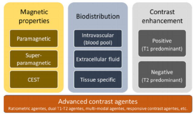
Despite the numerous families of contrast agents, they can be classified, in general, based on three different criteria:
- The magnetic properties of the contrast agents
- The in vivo biodistribution behavior of the contrast agent
- The type of contrast enhancement produced by the agent
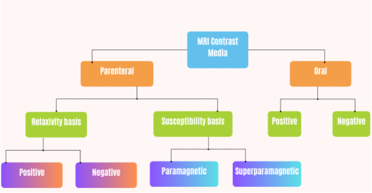
Most MR contrast agents used in human clinical applications are based on paramagnetic substances which are metal ions with unpaired electrons in the outer orbital shells (transition and lanthanide metal ions), giving rise to magnetic dipoles when exposed to an external magnetic field. Many paramagnetic metal ions could be used as MR contrast agents, but gadolinium or manganese ions (linked to chelating molecules of different nature and functionality) are the most used ones because of the seven unpaired electrons of Gd3+ ions (five for manganese), combined with long electron spin relaxation times.
Paramagnetic contrast agents alter the appearance of an image typically by shortening the T1 or T2 of water protons. Although Gd3+-based agents are typically referred to T1 agents because they have the biggest impact on the T1 of water protons, they certainly impact both T1 and T2 Solvent water molecules. The presence of a Gd3+-based contrast agent can be described at a minimum as three types:
- A single inner-sphere water molecule that binds weakly with the Gd3+ion and exchanges rather rapidly with all other nearby water molecules.
- A number of second-sphere water molecules that interact weakly with the entire Gd3+-complex.
- The remaining outer-sphere or bulk water molecules.
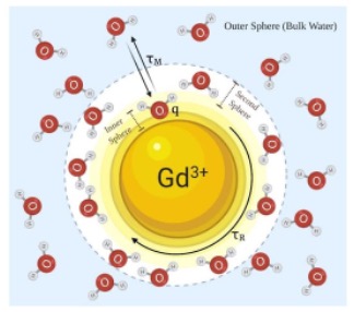
All protons on all water molecules are influenced by the presence of a Gd3+ complex in solution, the largest impact is felt by the single exchanging inner-sphere water molecule. This single water molecule, closest to the paramagnetic center, is quickly relaxed by the seven unpaired electrons on the Gd3+ and then dissociates from the inner-sphere position and is replaced by another nearby water molecule. The partially oriented water molecules in the second-sphere are also partially relaxed by the nearby Gd3+ ion, but they are further away from the paramagnetic center, so the relaxation efficiency is about 50% less than the single inner-sphere water molecule. If water exchange is rapid, then the entire sample of water molecules is impacted by the presence of Gd3+, and this relaxation effect translates into a brighter signal intensity in T1-weighted proton images.
Gadolinium enhances vasculature or pathologically-vascular tissues (e.g. intracranial metastases, meningiomas). This process involves injecting 5-15ml of contrast intravenously, with images taken shortly thereafter. Gadolinium appears bright in signal, allowing for detection of detailed abnormalities (e.g. intracranial pathologies).
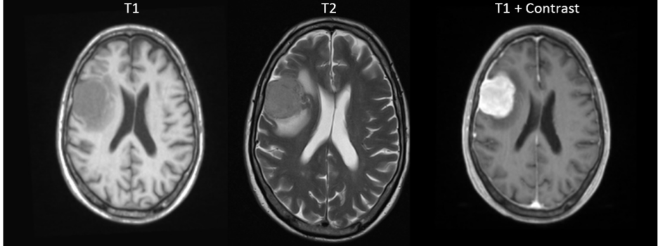
Meningioma is shown more clearly by gadolinium contrast
Super-paramagnetic agents are based on iron oxides, mostly magnetite (Fe3O4) or maghemite (γ-Fe2O3). These are water-dispersible crystals with a core diameter in the range 5−10 nm, made of several thousands of iron nuclei that are usually coated by a polymeric shell. Super-paramagnetic agents strongly reduce both T1 and T2 relaxation times of surrounding water molecules, but their dominant effect is on the transverse relaxation times (T2orT2*). Thus, super-paramagnetic contrast agents are usually referred as T2 contrast agents.
Gadolinium based Contrast Agents (GBCAs):
Regarding the chelate type and charge, GBCAs can be divided into linear/macrocyclic and ionic/non-ionic groups. A wide range of GBCAs are commercially available and their stability is dependent on the conditional thermodynamic stability constant (Kcond), thermodynamic stability constant (Ktherm), and kinetic stability.
Multiple studies show that the brain holds onto more gadolinium particles from linear agents than macrocyclic agents. Early studies show that most macrocyclic molecules filter through the kidneys and leave the body within 24 hours after an MRI. The rest exit the body within 72 hours.
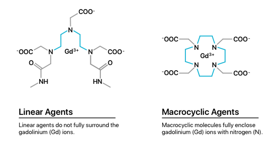
Ionic GBCAs are more stable than nonionic ones and the stability of macrocyclic compounds is higher than linear compounds. Hence, ionic macrocyclic agents are the most stable Gd chelates. Macrocyclic molecules bind strongly to Gd in an organized rigid ring; however, linear nonionic GBCAs have open chains and weaker binding to Gd. Compared to linear agents, macrocyclic agents are more stable in vivo. Low-stability GBCAs (linear, nonionic compounds) likely undergo transmetallation, release free Gd that deposits in tissues, attract fibrocytes, and therefore initiate the process of fibrosis.

Identification of the different GBCAs
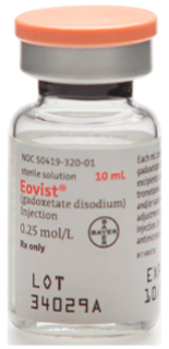
Eovist (gadoxetate disodium) is indicated for intravenous use in MRI of the liver to detect and characterize lesions in patients with known or suspected focal liver disease.
Magnevist (gadopentetate dimeglumine) Injection is a contrast agent that is used in combination with MRI to allow blood vessels, organs, and other non-bony tissues to be seen more clearly on the MRI used to help diagnose certain disorders of the heart, brain, blood vessels, and spinal tissues.

MultiHance (gadobenate) is a gadolinium based MRI contrast agent that has twice the relaxivity of conventional extracellular fluid (ECF) contrast agents, providing a marked increase in SNR to better visualize smaller lesions and improve the delineation of larger lesions.
In adult patients, MultiHance is indicated in Europe, and in several countries in Asia and Latin America, for MRI exams of the CNS (brain and spine) and for MRI of the liver, as well as for MRA (Magnetic Resonance Angiography), where it improves the diagnostic accuracy for detecting clinically significant steno-occlusive vascular disease in adult patients with suspected or known disease of the abdominal or peripheral arteries. In the US, MultiHance is indicated for MRI of the CNS, and it has recently received approval of the FDA for contrast-enhanced MR Angiography (MRA). In this indication, MultiHance can be used to evaluate adults with known or suspected renal or aorto-ilio-femoral occlusive vascular disease.

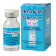
Dotarem (Gadoterate meglumine also known as gadoteric acid) is a contrast agent that has magnetic properties. It is used in combination with MRI to allow blood vessels, organs, and other non-bony tissues to be seen more clearly on the MRI. Dotarem is used to help diagnose certain disorders of the brain and spine (central nervous system).
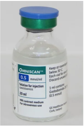
Omniscan (Gadodiamide) is indicated as a contrast medium for cranial and spinal MRI and for general MRI of the body after intravenous administration. Omniscan provides contrast enhancement and facilitates visualisation of abnormal structures or lesions in various parts of the body including the CNS.
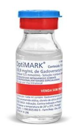
Optimark (gadoversetamide) Injection is indicated for use with MRI in patients with abnormal blood brain barrier or abnormal vascularity of the brain, spine and associated tissues. Optimark Injection is indicated for use with MRI to provide contrast enhancement and facilitate visualization of lesions with abnormal vascularity in the liver in patients who are highly suspect for liver structural abnormalities on computed tomography.
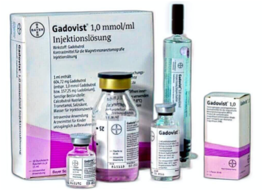
Gadovist (gadobutrol) injection is indicated in adults and children of all ages including term newborns for: Contrast enhancement during cranial and spinal MRI investigations and for contrast-enhanced magnetic resonance angiography (CE-MRA), Contrast enhanced MRI of the breast to assess the presence and extent of malignant breast disease, and MRI of the kidney.
Gadovist is particularly suited for cases where the exclusion or demonstration of additional pathology may influence the choice of therapy or patient management, for detection of very small lesions and for visualization of tumors that do not readily take up contrast media. Gadovist is also suited for perfusion studies for the diagnosis of stroke, detection of focal cerebral ischemia and tumor perfusion.
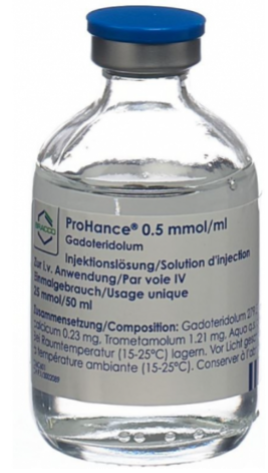
ProHance (Gadoteridol) Injection is indicated for use in MRI in adults and children over 2 years of age to visualize lesions with abnormal vascularity in the brain (intracranial lesions), spine and associated tissues. ProHance is indicated for use in MRI in adults to visualize lesions in the head and neck.
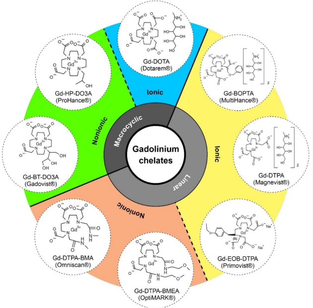
Structure of currently marketed GBCAs used for MRI.
Side effects of GBCA:
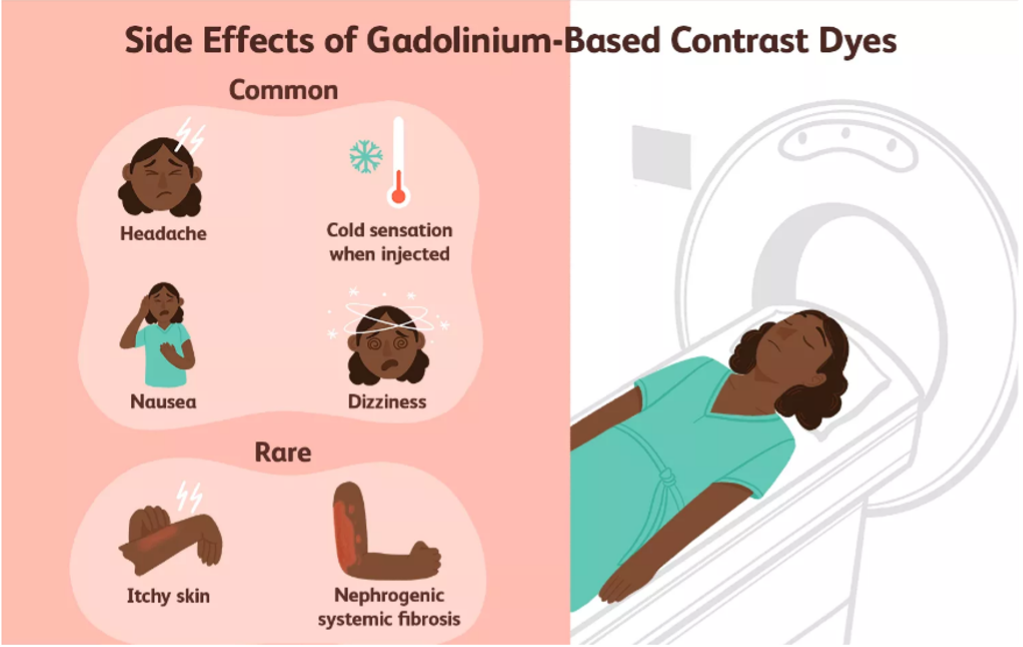
The dye frequently causes side effects, such as nausea, headaches, dizziness, and rash, but they tend to be mild. After an MRI, most GBCAs is removed from the body in urine, but trace amounts may stay in the body and accumulate over time in people who have MRI scans with GBCAs frequently.
Rarely, certain types of gadolinium contrast cause a severe disease called nephrogenic systemic fibrosis (NSF) in people with significant kidney dysfunction. This condition, which causes tightening of the skin and damage to internal organs, is most likely to occur in people with MS who also have kidney dysfunction. Although rare, some people have an allergic reaction to gadolinium contrast. The main symptom is itchy skin, but rashes have also been reported. A life-threatening allergic reaction, known as anaphylaxis, is also possible but unlikely.
Key Players:
Some of the prominent players operating in the Contrast Agents in MRI market are:
- Unijules Life Sciences Ltd (India)
- Spago Nanomedical AB (Sweden)
- Lantheus Medical Imaging Inc (U.S.)
- JB. Chemicals and Pharmaceuticals Limited (India)
- Guerbet LLC (France)
- GE Healthcare (U.S.)
- Daiichi Sankyo Company Limited (Japan)
- Bracco Imaging S.p.A (Italy)
- Bayer Healthcare AG (Germany)
- Jodas Expoim (India)
Resources:
https://www.mdpi.com/1424-8247/13/10/268
https://www.drugwatch.com/gadolinium/side-effects/
https://www.mdpi.com/2076-3263/9/2/93
https://www.verywellhealth.com/contrast-dyes-for-mri-in-ms-3972534
https://www.linkedin.com/pulse/contrast-agents-mri-market-analysis-detailed-outlook-forecast-smith/
https://geekymedics.com/the-basics-of-mri-interpretation/


